Using the origami technique to design RNA nanostructures
-
 Loading...
 - Ryan Taylor
- 01 Mar 2023
- 63 Views
- 0 Like
- 0 Comment
Researchers from Aarhus University and Berkeley Laboratory have designed RNA molecules, that folds into nanoscale rectangles, cylinders, and satellites, and have studied their 3D structure and dynamics with advanced nanotechnological methods. In an article in the journal Nature Nanotechnology, the researchers describe their work and how it has led to the discovery of rules and mechanisms for RNA folding that will make it possible to build more ideal and functional RNA particles for use in RNA-based medicine.
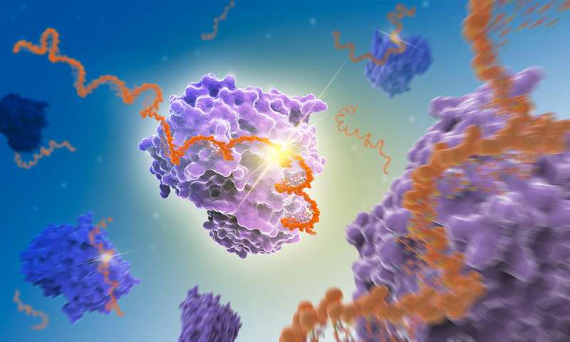
The RNA molecule is commonly recognized as messenger between DNA and protein, but it can also be folded into intricate molecular machines. An example of a naturally occurring RNA machine is the ribosome, that functions as a protein factory in all cells. Inspired by natural RNA machines, researchers at the Interdisciplinary Nanoscience Center (iNANO) have developed a method called "RNA origami", which makes it possible to design artificial RNA nanostructures that fold from a single stand of RNA. The method is inspired by the Japanese paper folding art, origami, where a single piece of paper can be folded into a given shape, such as a paper bird.
Frozen folds provide new insight
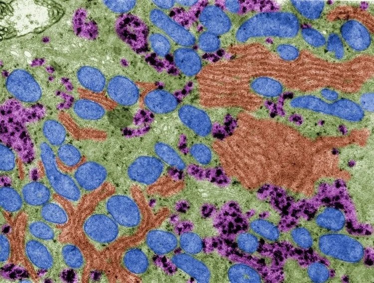
The research paper in Nature Nanotechnology describes how the RNA origami technique was used to design RNA nanostructures, that were characterized by cryo-electron microscopy (cryo-EM) at the Danish National cryo-EM Facility EMBION. Cryo-EM is a method for determining the 3D structure of biomolecules, which works by freezing the sample so quickly that water does not have time to form ice crystals, which means that frozen biomolecules can be observed more clearly with the electron microscope. Images of many thousands of molecules can be converted on the computer into a 3D map, that is used to build an atomic model of the molecule. The cryo-EM investigations provided valuable insight into the detailed structure of the RNA origamis, which allowed optimization of the design process and resulted in more ideal shapes.
Discovery of a slow folding trap
Cryo-EM images of an RNA cylinder sample turned out to contain two very different shapes, and by freezing the sample at different times it was evident that a transition between the two shapes was taking place. Using the technique of small-angle X-ray scattering (SAXS), where the samples are not frozen, the researchers were able to observe this transition in real time and found that the folding transition occurred after approx. 10 hours. The researchers had discovered a so-called "folding trap" where the RNA gets trapped during transcription and only later gets released (see video). Read More…