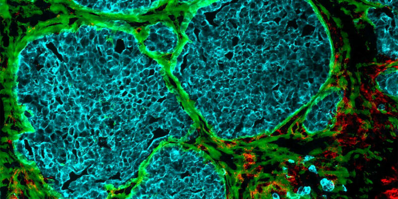
How tumours transform blood vessels
-
 Loading...
 - paula summer
- 17 Mar 2023
- 62 Views
- 0 Like
- 0 Comment
Increasingly dense cell clusters in growing tumours convert blood vessels into fibre-filled channels. This makes immune cells less effective, as findings by researchers from ETH Zurich and the University of Strasbourg suggest.
It was almost ten years ago that researchers first observed that tumours occurring in different cancers - including colorectal cancer, breast cancer and melanoma - exhibit channels leading from the surface to the inside of the cell cluster. But how these channels form, and what functions they perform, long remained a mystery.
Elaborate and detailed experiments
Through a series of elaborate and detailed experiments, the research groups led by Viola Vogel, Professor of Applied Mechanobiology at ETH Zurich, and Gertraud Orend from the University of Strasbourg have found possible answers to these questions. There is a great deal of evidence to suggest that these channels, which the researchers have dubbed tumour tracks, were once blood vessels.
These blood vessels start out by supplying the fast-growing cell clusters with glucose and oxygen. But then the vessels undergo a process that strips them of their original function of transporting blood: the vessel walls change and the vessel cavity gradually fills up.
When fibres control the behaviour of immune cells
This filler material consists mainly of cells and newly formed protein fibres, which make up what is known as the extracellular matrix. Collagen fibres are found here, as are fibronectin fibres. The latter play a role in growth processes that take place mainly during embryonic development or wound healing. In their external page article call_made , the researchers show that the fibres within the tumour tracks are capable of trapping immune cells. Read More…