Matching form and function of brain cell types
-
 Loading...
 - Yovana Molina
- 28 Apr 2023
- 70 Views
- 0 Like
- 0 Comment
Investigators at Cedars-Sinai have created computer-generated models to bridge the gap between "test tube" data about neurons and the function of those cells in the living brain. Their study, published in the journal Nature Communications, could help in the development of treatments for neurological diseases and disorders that target specific neuron types based on their roles.
This work allows us to start looking at the brain like the complex machine that it is, rather than as one homogenous piece of tissue," said Costas Anastassiou, Ph.D., a research scientist in the departments of Neurology, Neurosurgery and Biomedical Sciences at Cedars-Sinai and senior author of the study. "Once we are able to distinguish between the different cell types, instead of saying that the entire brain has a disease, we can ask which neuron types are affected by the disease and tailor treatments to those neurons."
Neurons are the main functional units of the brain. The signals passing through these cells—in the form of electrical waves—give rise to all thought, sensation, movement, memory and emotion.
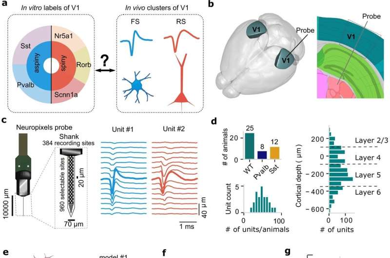
The study used data from laboratory mice to establish a new method for examining relationships between neuron type and function, and focused on the mouse primary visual cortex, which receives and processes visual information. It is one of the best studied parts of the brain—both in vitro, where tissue is studied in a dish or test tube outside the living organism, and in vivo, where it is studied in the living animal. Read More..